

Can You Live a Normal Life After Hernia Surgery?
Patient information prepared for Dr (Major) RS Rengan
A large hernia can look straightforward from the outside. There is a swelling, a scar and perhaps discomfort when standing or walking. Inside the abdomen, however, the picture may be very different. There may be several openings along an old incision, muscles pulled away from the midline, bowel within the hernia and mesh from an earlier operation.
This is why planning complex hernia surgery often begins well before entering the operating theatre.
For patients considering abdominal wall reconstruction in Chennai, understanding the CT scan can make the consultation more useful. It helps answer a practical question: what will it take to repair this particular abdominal wall?
Dr (Major) RS Rengan brings more than 37 years of surgical experience to the assessment of hernia and abdominal wall conditions. For a large, recurrent or long-standing hernia, the discussion should go beyond choosing between open and keyhole surgery. It should explain the anatomy, the likely reconstruction and the preparation needed. Dr (Major) RS Rengan: Professional profile
Why Examination Alone May Not Be Enough
Clinical examination remains essential. A surgeon assesses the swelling, skin, scars, tenderness and whether the hernia reduces when you lie down. A CT scan adds a cross-sectional view of structures that cannot be assessed fully through the skin.
The European Hernia Society recommends CT or MRI for preoperative planning when surgery for an incisional hernia is being considered. European Hernia Society: Midline incisional hernia guidelines
An incisional hernia develops through the scar of an earlier abdominal operation. A recurrent hernia has returned after a previous hernia repair. These categories can overlap.
What Does a CT Scan Show Before Hernia Surgery?
A useful CT assessment describes the abdominal wall as a whole. It can identify the number and dimensions of defects, the contents of the hernia sac and the surrounding muscles. It may also identify associated problems that change the surgical plan. Computed tomography for ventral hernia: Need for a standardised reporting format
The visible bulge and the opening beneath it are different measurements. A large sac may communicate with the abdomen through a smaller opening. Conversely, a long scar may contain several separate defects, sometimes described as a “Swiss-cheese” pattern.
For the patient, this explains why the final repair may need to cover a broader area than the swelling initially suggests.
Hernia Volumetry: Measuring How Much Lies Outside the Abdominal Cavity
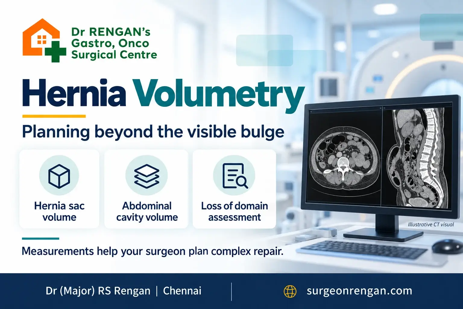
In a very large, long-standing hernia, a substantial amount of the abdominal contents may sit within the hernia sac. Hernia volumetry measures the sac and the remaining abdominal cavity to describe their relationship.
How Are Hernia Volumes Calculated?
The original Tanaka approach estimates volumes from CT dimensions. Modern segmentation methods outline structures across CT slices to obtain three-dimensional measurements. Tanaka et al.: CT-based hernia sac and abdominal cavity volume calculation, Deep learning in ventral hernia imaging
Tanaka Ratio and Sabbagh Ratio
Two ratios are commonly discussed:
| Measurement | Calculation |
|---|---|
| Tanaka ratio | Hernia sac volume ÷ remaining abdominal cavity volume |
| Sabbagh ratio | Hernia sac volume ÷ (hernia sac volume + remaining abdominal cavity volume) |
These ratios have different denominators and must not be used interchangeably. Preoperative algorithm for loss of domain hernia repair
For example, if a hernia sac measures 2 litres and the remaining abdominal cavity measures 8 litres, the Tanaka ratio is 25%, while the Sabbagh ratio is 20%. These describe the same anatomy using different calculations. They do not mean a 25% or 20% chance of surgical failure.
What Is Loss of Domain?
“Loss of domain” describes a situation in which returning the herniated contents and closing the abdominal wall may be difficult or physiologically poorly tolerated. Volumetry helps assess this problem, although it cannot capture every aspect of abdominal wall flexibility or a patient’s breathing reserve.
A Tanaka ratio around 25% is commonly used as a planning flag in large hernias. It is not an automatic instruction to perform one particular operation. Tanaka et al.: CT-based volumetry, Preoperative algorithm for loss of domain hernia repair
Preparing Patients with Loss of Domain
In selected patients, specialist preparation may include botulinum toxin injections to relax the lateral abdominal muscles or progressive pneumoperitoneum to gradually increase abdominal capacity. These measures require individual assessment and are not routine for every large hernia. Preoperative algorithm for loss of domain hernia repair
Previous Mesh: Why the Earlier Operation Matters
If you have already undergone hernia repair, the surgeon needs to understand what was done previously. Relevant questions include where the mesh was placed, how far it extends and where the new defect lies in relation to it.
What Can CT Reveal About Previous Mesh?
CT may help identify mesh, fixation devices, surrounding collections and the relationship between the repair and nearby bowel. However, some mesh materials are difficult to see, and the precise mesh plane may remain uncertain. Computed tomography for ventral hernia: Need for a standardised reporting format
Why Previous Surgical Records Matter
A CT scan cannot reliably map every adhesion or always establish whether mesh is infected. The scan must be interpreted alongside symptoms, examination and previous operative records.
Bring your earlier discharge summary, operation note and mesh details if available. These records may answer questions that imaging cannot. A recurrent hernia does not, by itself, establish that every piece of old mesh must be removed; this needs a specific discussion with the operating surgeon.
EHS Classification: Giving the Hernia a Precise Description
The European Hernia Society (EHS) classification describes incisional hernias by location, width and recurrence status. It helps clinicians communicate clearly and compare similar hernias in research. European Hernia Society: Classification of primary and incisional abdominal wall hernias
Midline Hernia Locations: M1 to M5
| Code | Location |
|---|---|
| M1 | Subxiphoidal, just below the breastbone |
| M2 | Epigastric, in the upper central abdomen |
| M3 | Umbilical, around the navel |
| M4 | Infraumbilical, below the navel |
| M5 | Suprapubic, just above the pubic bone |
Lateral Hernia Locations and Width Categories
Lateral defects use L1 to L4 for subcostal, flank, iliac and lumbar locations. A hernia can span more than one region.
The commonly used width groups are W1: less than 4 cm; W2: 4–10 cm; and W3: greater than 10 cm. The original classification contains overlapping wording at the 10 cm boundary, so the exact measured width should always accompany the code.
The classification is an anatomical description. It is not a cancer stage or a stand-alone prediction of risk. Two patients with the same category may still require different operations because their muscles, previous repairs and general health differ.
Can CT Predict Whether Component Separation Will Be Needed?
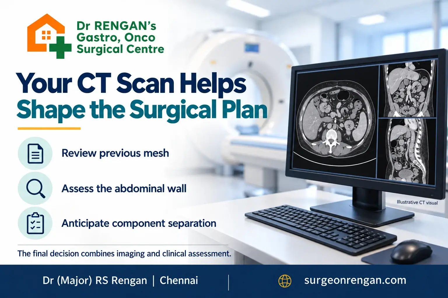
CT can indicate that closing the abdominal wall is likely to require additional release. It cannot determine the operation with certainty.
CT Findings Associated with Myofascial Release
For midline incisional hernias, the EHS guideline identifies several findings associated with a greater likelihood of needing myofascial release:
- A defect width greater than 8 cm.
- A defect area greater than 164 cm².
- A rectus-to-defect ratio below 1.34.
- A component separation index above 0.146.
European Hernia Society: Midline incisional hernia guidelines
These are planning indicators drawn from limited evidence. They should not be treated as universal cut-offs. The rectus-to-defect ratio relates the combined rectus muscle widths to the gap; the component separation index is a specialised CT measure of the separation geometry.
What Is Component Separation?
Component separation releases selected abdominal wall layers so that tissues can move towards the midline. The objective is to help reconstruct the wall without forcing widely separated edges together under excessive tension.
Anterior Component Separation and Posterior Component Separation with TAR
Options include anterior component separation and posterior component separation with transversus abdominis release (TAR). TAR can provide additional movement and space for a broad mesh behind the muscles.
“Component separation” does not mean removing the abdominal muscles. It describes a planned release based on their anatomy. American College of Surgeons: Contemporary management of complex abdominal wall hernias
Can the Scan Help Choose Open, Laparoscopic or Robotic Surgery?
CT contributes to that decision, but an imaging measurement alone cannot select the best approach. Defect geometry, previous mesh, available tissue planes and the extent of reconstruction all enter the discussion.
Questions to Ask About Your Surgical Approach
Ask your surgeon to explain the proposed operation in practical terms:
- Will the abdominal wall defect be closed?
- Where is the mesh likely to sit?
- Is a muscle release anticipated?
- What findings during surgery could change the plan?
These questions are more informative than choosing an approach solely because it uses smaller incisions. The goal is a reconstruction suited to your anatomy, health and priorities.
Can a CT Scan Predict Recurrence or Complications?
What CT Measurements Can and Cannot Predict
Anatomical planning and outcome prediction are different tasks. CT can describe the hernia well without reliably predicting whether it will recur.
A study of 188 patients found that most preoperative CT measurements, including loss-of-domain ratios, did not distinguish those with recurrence at one year from those without it. Study of preoperative CT variables and ventral hernia repair outcomes
Why Overall Health Still Matters
Risk assessment therefore needs more than an image. Patient health, smoking, diabetes, fitness, previous complications and the reconstruction itself all matter. A detailed scan supports a clinical decision; it does not provide a guarantee.
Dr Vinayak Rengan, Curium Life and AI in Hernia Planning
Dr RS Rengan’s son, Dr Vinayak Rengan, is a paediatric and neonatal surgeon and the founder of Curium Life, a surgical AI company developing tools to support surgical assessment and decision-making.
AI-Assisted Hernia Assessment
His work includes AI-assisted hernia assessment, with the broader aim of combining imaging and clinical information to help predict surgical complexity and outcomes. Potential applications include automating volumetry, assessing abdominal muscles and producing more consistent measurements for surgical planning.
Deep Learning Research in Ventral Hernia Imaging
A 2026 research paper led by Dr Vinayak Rengan analysed 215 adult ventral hernia CT scans using deep learning to outline hernia defects and relevant muscles. Curium Life supported algorithm development, training and testing. Deep learning in ventral hernia imaging: Automated multi-structure CT segmentation for surgical planning
This is an encouraging foundation, but the distinction matters: identifying structures accurately on a CT scan is not the same as proving that AI can predict an individual patient’s recurrence or prevent complications. The published study describes a proof of concept requiring prospective clinical validation before use in operative decision-making.
For patients, the purpose of this research is to make complex information easier for the surgical team to measure and interpret, while keeping clinical judgement central.
Questions Patients Often Ask
Q: Does every hernia need a CT scan?
A: No. Some straightforward hernias can be assessed clinically. Imaging becomes particularly useful when the diagnosis is uncertain or the anatomy needs detailed evaluation before incisional hernia repair. European Hernia Society: Midline incisional hernia guidelines
Q: Does the scan always require contrast?
A: No. The protocol depends on the clinical question. Contrast may be useful when assessing bowel or suspected complications. Tell the team about kidney disease, previous contrast reactions and any possibility of pregnancy before scanning. Computed tomography for ventral hernia: Need for a standardised reporting format
Q: Should I bring the images or only the report?
A: Bring both if available. The original digital images, ideally in DICOM format, allow the surgeon to review the anatomy and measurements directly. Also bring records of previous abdominal operations.
Q: Does a large hernia automatically require TAR?
A: No. Size is one consideration. Muscle position, tissue quality, previous surgery and the ability to close the defect also influence whether a release is needed.
Q: Can AI tell me exactly which operation I need?
A: No. Current research supports the feasibility of automated measurements. An individual surgical recommendation still requires examination, clinical assessment and specialist interpretation. Deep learning in ventral hernia imaging
Discuss Your Complex Hernia with Dr RS Rengan in Chennai
If you have a large abdominal hernia, a recurrence after mesh repair or a swelling that has grown over many years, bring your previous records and any existing CT images to your consultation.
A useful consultation should leave you understanding what the scan shows, why a particular reconstruction is being considered and what preparation may help before surgery.
Book a consultation with Dr (Major) RS Rengan to discuss your hernia and an individualised treatment plan.
Sudden severe pain, repeated vomiting, a newly irreducible swelling or skin discolouration over a hernia requires urgent medical assessment rather than waiting for an elective appointment.
Berlin Sharmila
Senior Content Editor Healthcare SEO Specialist
Berlin Sharmila is a Senior Content Editor with expertise in healthcare content, digital marketing, and SEO. She creates clear, patient-focused content that helps readers make informed medical decisions.
Related posts


